Causes de dystocie chez les brebis de la région de Djelfa (Algérie)
Résumé
La présente étude a comme objectif de déterminer les causes de dystocie chez les brebis dans la région de Djelfa (Algérie). 87 brebis dystociques ont fait l’objet de cette étude. Les causes de dystocie fœtale étaient plus fréquents (75 %) que les causes maternelles (16 %). Les causes materno-fœtales représentaient 9 %. Plusieurs origines de la dystocie ont été enregistrées. La malprésentation était la cause la plus importante de dystocie, suivie de l’emphysème, de la non dilatation du col utérin et de l’inertie utérine. Les autres causes étaient moins importantes. En ce qui concerne l’effet du type de dystocie sur la viabilité des agneaux, nous avons constaté que le grand nombre d’agneaux morts avait été observé en cas d’emphysème fœtal.
Mots-clés: Algérie, Brebis, Djelfa, Dystocie
Téléchargements
INTRODUCTION
Small ruminant production is one of the main sources of meat production (≈31 millions heads) in Algeria, which plays a vital role in food security. The small ruminant population in Algeria stands as 27 millions sheep and 4 millions goats respectively, where 65 % of the total populations are females and 35% males. However, both sheep and goats are reared under traditional extensive system in Algeria, intensive husbandry systems were recently been introduced in the country (MADR 2014; Kardjadj et al., 2016).
In spite of the population advantage of small ruminants, diseases and poor herd-health management practices poses a significant challenge to efficiently and profitably manage small ruminants’ production in developing world such as Algeria. However, the viability of sheep and goat farming depends largely on their reproductive performance that is invariably regulated by genetic and environmental factors (Mellado et al., 2006; Kardjadj et al., 2016).
Abnormal or difficulty in giving birth is referred to as dystocia (Youngquist et al., 2007). Blood et al. (2011) also defined dystocia as difficulty in parturition to the point of needing human intervention.
In small ruminants, dystocia or difficult birth contribute to significant economic loss in terms of loss of perinatal death of dams and fetus, uterine infections, more retained placentas, and longer lambing and kidding intervals (Rook et al., 1990; Ghosh et al., 1992; Brounts et al 2004; Scott 2005).
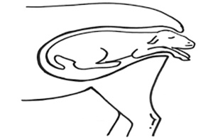
Generally, dystocia may be of fetal or maternal origin (Noakeset al.,2009; Ali 2011). Fetal causes of dystocia include mainly oversize, maldisposition and monsters (Majeed and Taha 1989a; Noakes et al.,2009). Maternal causes of dystocia include mainly incomplete cervical dilatation (ringwomb), narrow pelvis, and uterine inertia (Majeed and Taha 1989b; Thomas 1992; Noakes et al., 2009).
The incidence of dystocia is generally influenced by factors such as breed of sire, breed of dam, age of dam, number of fetus and body weight of dam (Hanie, 2006). As dystocia is usually considered a major cause of newborns and dams deaths with severe economic losses, thus, the aim of this study was to describe the common causes of dystocia in small ruminants reared in Ain Ouassara in the wilaya of Djelfa, a semi-arid area.
MATERIALS AND METHODS
Study area
The wilaya of Djelfa is located in the high plateau, south of Medea, 300 kilometers south of Algiers with an area of 256.35 km2. Its continental climate is marked by cold, wet winters and hot dry summers. However, landscapes and landforms are relatively varied. Forests occupy 8 % of the territory. It is a pastoral-oriented department where steppe predominates and sheep are the most numerous with a total of more than 4 million heads. Djelfa market is one of the major markets for Algeria sheep. The Wilaya has a population of more than 1,164,000 inhabitants, mainly concentrated in the cities of Djelfa, Ain Oussara, Messaadi and Hassi Bahbah (Hamrat et al., 2011). The present study was carried out in Ain Ouassara region.
Study design
A total of eighty seven sheep suffering from dystocia and presented to a private veterinary clinic in the wilaya of Djelfa province were the subject of this study from September 2017 to March 2018. Immediately after presentation, anamnesis and clinical status of each animal was recorded. Dystocia was considered when the mothers have more than one hr of active labour without producing a newborn (Bowen, 1978; Ali, 2011).
Species, age, parity of the dam, date of presentation, viability and number of newborn were recorded. Different etiological causes of dystocia were also recorded.
RESULTS
Some 87 cases of dystocia were recorded in sheep during the period of the study. Fetal causes of dystocia were more prevalent 75 % (65/87) than maternal causes 16 % (14/87). Materno-fetal causes represented 9 % (8/87).
Malpresentation was the most important cause of dystocia, followed by emphysema, ringwomb and uterine-inertia. Others causes were less important (Table 1).
In the present study, fetal and maternal dystocia occurred more commonly in multiparious (Table 2).
Also, 62 single litters, 22 double litters and 3 triple litters were recorded in sheep. Dystocia was strongly associated with single litters size, followed by double litters size (Table 3).
In regards to effect of dystocia type on the viability of lambs, we have recorded that the large number of dead lambs was observed in case of fetal emphysema (Table 4).
DISCUSSION
Dystocia is often a major cause of lamb loss in the flock and may result in great economic loss to the farmers (Faraidoon and Talib, 2010). The preponderance of dystocia in sheep over other domestic ruminants has also been reported in other study (Ahmed et al., 2017).
Dystocia is naturally regarded as being either of maternal or fetal origin (Ahmed et al., 2017). During the present study fetal origin was the common. This result is in agreement with reports by Ahmed et al. (2017) but is in contrast with the result of Ali (2011) who reported more of maternal causes of dystocia in Saudi Arabia.
Our results showed that fetal dystocia occurred mainly due to malpresentation, which is in agreement with the results of Ahmed et al. (2017).
Fetal emphysema represented second fetal origin with 13.8 %. Kumar et al. (2013) reported 4 cases of fatal emphysema, representing 23.5% from the 17 small ruminants studied.
In the present study, we have reported other less important fetal cases such maceration (1.15 %), malformation (1.15%) and anasarc (10.1 %). Fetal anasarc has been reported in Awassi sheep newborns by Hailat et al. (1997). Fetal malformation (1.15 %) was observed as a cause of dystocia in sheep in this study. Based on studies in Australia, New Zealand and USA, the incidence of congenital defects ranges from 0.2 to 2.0 % of all lambs born (Dennis and Leipold, 1979). The important incriminated factors are prenatal viral infection, intrauterine exposure to poisons ingested by the dam, vitamin deficiency like vitamin A and folic acid, hyperthermia, and gene mutation (Jones et al., 1997; Ali, 2011).
From the maternal origin, ringwomb was the third cause. Cases of ringwomb have been also reported by Noakes et al. (1996); Ahmed et al. (2017). The incomplete cervical dilatation was observed as the most common cause of dystocia in sheep and goats by Kumar et al. (2013). Failure of the cervix to dilate may be attributed to failure of secretion of the hormones that control labor or of the tissue response to hormonal secretion (Wu et al., 2004; Palliser et al., 2006 et Ali, 2011). 4.6 % of the maternal cases were represented by uterine torsion. This result corroborates with 4.4 % reported by Ali 2011.
During this study, maternal-fetal dystocia was the most frequent in primiparuous (62.5 %), followed by maternal dystocia (35.7 %). Ali (2011) reported that maternal dystocia occurred more frequently in primiparuous. In multiparuous, fetal origin was more frequent than maternal dystocia (73.8 %). This situation corroborates with the finding of Ali (2011).
In regards to birth type or litter size, the frequency of dystocia was higher for singles than twins and triplets with 41.3. The explanation was given by Majeed et al. (1992) who found the same situation in goats and noted that the relative frequency of twins and triplets would lower the incidence of oversized fetuses and therefore reduce the rate of dystocia.
The present study showed that globally, the number of live newborns was very close to those died with 58 against 57. Contrary to what reported Bhattacharyya et al., (2015)which recorded a predominance of dead fetuses (60) in small ruminants against only 18 alive.
By origin, it may be noted that in the present study, fetal causes marked more deaths (47 versus 40) than living because the fetal origins were predominant (mainly malpresentations and emphysema).
CONCLUSION
Dystocia is a common problem in lambing ewes. It constitutes a major reproductive problem among small ruminants and can hinder or affect their productivity. We recommend that more practical and reliable means of handling such cases should be encouraged among veterinary personnel as well as the need for more training of such staff.
REFERENCES
Abdullah F.F.J., Chung E.L.T., Sdiq M.A., Abba Y., Tijjani A., Mohammed K., Osman A.Y., Laila M.A.M. (2015) Management of fetal dystocia caused by carpal flexion in ewe: a case report. J. of Advances Veterinary Animal Research, 2: 225-228.
Ali A.M.H. (2011). Causes and Management of Dystocia in Small Ruminants In Saudi Arabia. Journal of Agricultural and Veterinary Sciences. 4: 95-108
Blood D.C., Studdert V.P., Gay C.C. (2011). Saunders Comprehensive Veterinary Dictionary (4th Edition). London: Saunders.
Bowen J.S. (1978). Pregnancy toxemia, milk fever and kidding difficulties. Dairy Goat J., 56: 20-25.
Brounts S.H., Hawkins J.F., Baird A.N., Glickman L.T. (2004). Outcome and subsequent fertility of sheep and goats undergoing cesarean section because of dystocia: 110 cases (1981-2001). Journal of the American Veterinary Medical Association, 224: 275-281.
Dennis S.M., Leipold H.W. (1979). Ovine Congenital Defects. The Veterinary Bulletin, 49: 233 237.
Faraidoon A.M.A., Talib G.M.A. (2010). Treatments of dystocia in Karadi ewes in Sulaimani Province. Bas.J.Vet.Res., 9: 35-39.
Ghosh A., Yeasmina F., Alam M.G.S. (1992). Studies of ringwomb in Black Bengal goats. Theriogenology, 37: 527-532.
Hailat N., Lafi S.Q., al-Darraji A., El-Maghraby H.M., Al-Ani F., Fathalla M. (1997). Foetal anasarca in Awassi sheep. Aust. Vet. J., 75: 257-259.
Hamrat K., Achour Y., Ghadiri Y., Cozma V. (2011) Epidemiologic study of hydatidosis in the steppe regions of Djelfa, Algeria. Sci Parasitol., 12: 177-183.
Hanie E.A. (2006). Obstetrical Procedures. In: Large Animal Clinical Procedures for Veterinary Technicians. Elsevier, Mosby, Missouri. p. 413-31.
Jones T.C., Hunt R.D., King N.W. (1997). Veterinary pathology. T.C. Jones, R.D. Hunt, N.W. King (eds), William and Wilkins, Baltimore, USA.
Kardjadj M., Kouidri B., Metref D., Luka P.D., Ben-Mehdi M.H. (2015). Abortion and various associated risk factors in small ruminants in Algeria. PRE.VET. MED, http://dx.doi.org/10.1016/j.prevetmed. 2015.11.015
Kumar V., Talekar S.H., Ahmad R.A., Mathew D.D., Zama M.M.S. (2013). Delayed cases of dystocia in small ruminants - etiology and surgical management. Indian J. Vet. Sci., 1: 47-54.
MADR (2014). Ministère de l’Agriculture et de Développement Rural, Algérie. rapport Annuel.
Majeed A.F., Taha M.B. (1989a). Dystocia in local goats in Iraq. Small Ruminat Research, 2: 375-381.
Majeed A.F., Taha M.B. (1989b). Preliminary study on treatment of ringwomb in Iraqi goats. Animal Reproduction Science, 18: 199-203.
Majeed A.F., Taha M.B., Azawi O.I. (1992). Caprine Caesarean section. Small Ruminant Research, 9: 93-97.
Mellado M., Valdez R., Garcia J.E., Lopez R., Rodriguez A. (2006). Factors affecting the reproductive performance of goats under intensive conditions in a hot arid environment. Small Rumin. Res., 63: 110-118.
Noakes D.E., Parkinson T.J., England G.C.W. (2009) Arthuts’ Veterinary reproduction and obstetrics, 9th edition, England (eds), Saunders, Edinburg, London.
Palliser H.K., Hirst J.J., Gregory E., Rice G.E., Guck T., Ooi G.T., Dellios N.L., Escalona R.M., Ross Young I. (2006). Labor-Associated Regulation of Prostaglandin E and F Synthesis and Action in the Ovine Amnion and Cervix. J. Soci. Gyneco. Inves., 13: 19.
Rook J.S., Scholman G., Wing-Proctor S., Shea M. (1990). Diagnosis and control of neonatal losses in sheep. Veterinary Clinics of North America: Food Animal Practice, 6: 531-562.
Thomas J.O. (1992). Survey of the causes of dystocia in sheep. Veterinary Reccord, 127: 574-575.
Wu W., Xiao X., Hong M.A., CoksayganT., Chakrabarty K., Collins K.V., Rose J., Nathanielsz P.W. (2004). Prostaglandin mediates premature delivery in pregnant sheep induced by estradiol at 121 days of gestational age. Endocrinol., 45: 1444-1452. https://doi.org/10.1210/en.2003-1142PMid:14645114
Youngquist R.S., Threlfall W.R. (2007). Current therapy in large animal theriogenology (2nd Edition). Elsevier Health Sciences.
Téléchargements
Publié-e
Numéro
Rubrique
Licence

Revue Marocaine des Sciences Agronomiques et Vétérinaires est mis à disposition selon les termes de la licence Creative Commons Attribution - Pas d’Utilisation Commerciale - Partage dans les Mêmes Conditions 4.0 International.
Fondé(e) sur une œuvre à www.agrimaroc.org.
Les autorisations au-delà du champ de cette licence peuvent être obtenues à www.agrimaroc.org.