Analyse phylogénétique d'isolats du virus marocain de la clavelée basée sur le gène P32
Résumé
La clavelée (SP) est une maladie considérée hautement contagieuse par l’Organisation Mondiale de la santé Animale (OIE). L’agent causal de la maladie (SPPV) appartient au genre des Capripoxvirus contenant ainsi le virus de la variole de chèvre (GTPV) et le virus de la maladie nodulaire cutanée (LSDV). Le SPPV cause des pertes économiques considérables dans les zones endémiques telles que l’Afrique du nord et centrale, l’Asie, l’inde et le moyen orient. Au Maroc, peu d’étude de caractérisation moléculaire du SPPV sont disponibles, d’où l’objectif du présent travail qui vise à évaluer la relation génétique entre les souches virales marocaines isolées à partir de différentes régions du Maroc durant les flambées épizootiques, des souches vaccinales et des souches de références publiées sur Genbank, et ce, par le séquençage du gène P32. Toutes les séquences sont analysées par le logiciel MEGA 7.0, l’arbre phylogénétique est généré par la méthode Neighbour-Joining. Il apparaît clairement que tous les SPPV rapportés dans la plupart des pays sont branchés et groupés dans le clade du SPPV, et ont montré une forte relation génétique entre eux avec une identité d’acides nucléiques et d’acides aminés de 99-100 % et 98-100 % respectivement. Ces résultats nous mènent à conclure que le gène P32 apparaît hautement conservé chez tous les SPPV et les Capripoxvirus. Pour cela, plus d’études génétiques sont nécessaires afin de contrôler et expliquer la situation épidémiologique du SPPV.
Mots-clés: Sheeppox virus, gène P32, analyse phylogénétique, Capripoxvirus, Goatpox virus.
Téléchargements
INTRODUCTION
The Capripoxvirus genus, one of the eight members of the subfamily Chordopoxvirinae, is comprised of three important pathogens that infect only ungulates, Sheeppox virus (SPPV), Goatpox virus (GTPV) and Lumpy skin disease virus (LSDV), which are respectively causing disease in sheep, goats and cattle (Diallo and Viljoen, 2007).
Sheeppox, considered as notifiable animal diseases by the World Organization for Animal Health (OIE), is mainly endemic in central and northern Africa, the Middle East, India and other Asian countries from Central Asia to China (Diallo and Viljoen, 2007; Babiuk et al., 2008). SPPV is responsible for one of the most economically significant diseases of domestic ruminants in Africa and Asia (Carn, 1993; Esposito and Fenner, 2001). In Morocco, SPPV has been reported in enzootic form (CFSPH, 2017) and is still responsible for serious economic damage in sheep (FAO, 2010). It seems that the sheeppox disease appears only in sheep and no case affecting goats has been reported, which means that the entire circulated virus for several outbreaks in different regions of Morocco affects solely sheep (Zro et al., 2014).
Their genomes consist of double-stranded DNA of around 150 kb with terminal repeated sequences at each end. SPPV and GTPV genomes are very similar to that of lumpy skin disease virus (LSDV), sharing 97 % nucleotide identity (Tulman et al., 2002).
The usual criteria for classifying CaPVs is based upon the animal species from which the viruses are isolated, that is, SPPV from sheep, GTPV from goats and LSDV from cattle. But now, research has shown that some strains of SPPV and GTPV could infect both sheep and goats (Diallo and Viljoen, 2007; Bhanuprakash et al., 2010). Thus, strains identification based on the host animal species from which the strain was first isolated is not valid (Le Goff et al., 2009; Lamien et al., 2011).
Serological tests cannot differentiate SPPV from GTPV, because of the very close antigenic relationship among CaPVs (Balisky et al., 2008). So, the identification of these pathogens needs molecular methods. Fortunately, some molecular techniques for differentiation of CaPVs targeting specific genes had been developed, viz., the P32 gene (Hosamani et al., 2004), the RPO30 gene (Lamien et al., 2011), and the GPCR gene (Le Goff et al., 2009).
P32 is highly conserved among capripoxviruses. Its sequence information can them be used to differentiate SPPV, GTPV and LSDV, presenting the genetic relationship among different virus strains (Lamien et al., 2011; Hosamani et al., 2004). Moreover, it corresponds to an envelope protein homologous to P35 protein encoded by Vaccinia virus H3L gene and located on the membrane surface of the mature intracellular viral particle (Tulman et al., 2002).
This gene was targeted in this work, reported for the first time in Morocco, in order to carry out a phylogenetic analysis and to assess the genomic relatedness between Moroccan viral strains isolated from different geographic regions, vaccine strains and other reference strains retrieved from Genbank.
For this study, 24 Moroccan Sheeppox isolates mentioned above, 5 SPPV and GTPV vaccine strains were used. Furthermore, the sequencing results were used for phylogenetic analysis by comparing them with different capripoxvirus isolates retrieved from GenBank to evaluate the genetic relationship between them.
MATERIAL AND METHODS
Viruses
24 Cell-cultured strains isolated in Morocco from 1981 to 2010, 3 reference strains, 2 vaccine strains and 1 goat pox virulent strain were used for the study (Table 1).
These field isolates were compared to the corresponding sequences of 7 SPPV, 2 GTPV and 1 LSDV isolates retrieved from GenBank (Table 2).
DNA extraction, amplification and sequencing
DNA extraction
For viral DNA isolation from the samples, the Purelink viral DNA/RNA kit, INVITROGEN extraction kit was used according to the manufacturer’s instructions and eluted in 50 ml of elution buffer.
Polymerase chain reaction (PCR)
The pair of primers P32-1 (5’-ATG GCA GAT ATC CCA TT-3’) and P32-2 (5’-TTA CCA CAG GCT ATT AGA AG-3’) amplify the 1181 bp fragment containing the complete P32 ORF (Zhou et al., 2012) were used in PCR.
The PCR mixture contained 120 ng of extracted DNA, 5 μl5×PSbuffer, 0,2 μl Taq DNA polymerase 5U/ul, 8 μl 2.5 mM dNTP, 25 μl H2O, 1 μl of primer. DNA amplification was performed by ABI Verity Thermal Cycler. Thermal cycling parameters were: initial denaturation at 95°C for 1 min, then 35 cycles of: denaturation at 95°C for 15 sec, annealing at 49°C for 20 sec, and extension at 72°C for 3 min, followed by final extension at 72°C 20 min. The PCR product sizes were checked by the Qiaxcel bio analyzer, which generates virtual gels on the device software.
Sequencing
All obtained amplicons were purified using ExoSAP-IT treatment (USB Corporation, Cleveland, OH, USA) in order to achieve the Bi-directional sequencing which was performed by using a BigDye terminator cycle sequencing kit (Applied Biosystems, Foster City, CA, USA) and an ABI PRISM 3130XL automated DNA sequencer. Purified DNA was used at a concentration of 100 ng/μl for each sample and each reaction in a final volume of 10 μl. The thermal cycler program was: initial denaturation at 96°C for 1 minute and denaturation at 96°C for 10 seconds, annealing at 50°C for 5 seconds and 4 minutes of elongation at 60°C for 25 cycles.
Sequence alignment and phylogenetic analysis
For phylogenetic analysis, nucleotide sequences of related strains for P32 gene were retrieved from GenBank using online BLAST program on NCBI database. Multiple-alignment of these sequences was performed by MEGA 7 with ClustalW method. Once aligned, Neighbor-Joining tree (Saitou et al., 2016; Tamura et al., 2004) were constructed using MEGA 7 software (Kumar et al., 2016) with the Tamura 3-parameter model and Gamma distribution. The statistical significance of the nodes was assessed by bootstrap resembling analysis (1000 replicates) (Felsenstein, 1985). Sequences obtained in the current study were deposit in the GenBank database under the following accession numbers: MG201788 to MG201815 and KY769277 to KY769282.
RESULTS
Sequence analysis of P32 gene
The complete open reading frame (ORF) of P32 gene, of SPPV Moroccan isolates were sequenced and subjected to similarity analysis.
The results showed that all Moroccan SPPV samples shared a closed relationship between them, the nucleotide sequence identities varied between 91 and 100 %. The homology percentages of the amino acid sequence reached 91.5 to 100 % for Moroccan strains and varied from 98 to 100 % in comparison with the SPV reference strains.
Fortunately, BLAST results based on NCBI database showed that all of them had a high identity with SPPV strains (99-100 %) but the homology with GTPV and LSDV was 98 %.
Phylogenetic analysis
Using the phylogenetic analysis of the P32 gene performed on MEGA7, we concluded that CaPVs comprise 3 groups: GTPV, SPPV, and LSDV (Figure 1) and that SPPVs are more related to GTPVs than LSDV.
As shown, all of Moroccan isolates were clustered together and formed a single cluster with SPPVs strains isolated from distant countries like India, China, Russia, Iraq and Saudi Arabia; and from nearest countries like Tunisia. Furthermore, vaccine strains used in Morocco and Algeria were also clustered together with viral isolates.
According to the result obtained, Moroccan isolates are closely related despite the date of isolation and the geographical origins. Furthermore, we confirmed that the P32 gene is highly conserved region among the genome of capripoxviruses, the conservatism percentage per sites was 100% (Figure 2).
DISCUSSION
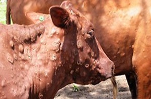
Sheeppox (SP) is an infectious viral disease, highly contagious, classified by the World Organization for Animal Health in the list of reportable diseases (Diallo and Viljoen, 2007). Clinically, sheeppox is characterized by hyperthermia and skin lesions (Hajjou et al., 2017). By its gravity, it is considered the most lethal animal pox. It develops either in a classic form (vesicular or nodular) or in a complicated form (Babiuk et al., 2008). It’s responsible of highest economic losses on meat and wool in Africa and worldwide (Diallo and Viljoen, 2007).
According to the animal health information published by the National Office for Food Safety, SP is enzootic in Morocco. Recently, 5 outbreaks (71 cases) were reported in the eastern and northern regions in January 2017 and caused the death of 12 affected animals. This situation leads us to understand why the disease still exists in Morocco despite vaccination programs and controls adopted by veterinary services.
In the present study, 24 isolates were collected from different regions of Morocco and spread over different years from 1981 to 2010 during several outbreaks. These isolates have undergone routine diagnostic tests, such as cell culture isolation and virus-neutralization (VNT) and enzyme-linked immunosorbent assay (ELISA), but these methods can not differentiate between SPPV, GTPV and LSDV. For this reason, and because of the few molecular studies of SPPV in Morocco, we opted for the sequencing of the viral envelope protein gene P32 of these isolates. Firstly, this gene was amplified by using polymerase chain reaction and sequenced to analyze the nucleotide divergence and to study the relationship between Moroccan isolates and other CaPVs strains retrieved from Genbank.
Sequencing of the P32 envelope protein gene, which is a structural protein containing the most important antigenic determinants and is present in all species of CaPVs (Heine et al., 1999; Tian et al., 2010), showed after nucleotide and protein sequence analysis that there exists a stronger relationship between the Moroccan strains of SPPV. They are clustered in the same clade of SPPV on the phylogenetic tree, with homology percentages ranging from 91 to 100 %. Furthermore, some restriction analysis of virus’s genomes isolated from different countries revealed that SPPVs are closely related (Black et al., 1986).
However, BLAST results on the NCBI database showed that all of local isolates of SPPV had a high identity (99-100 %) with SPPV strains but the homology with GTPV and LSDV was 98 %, which confirms the hypothesis that the GTPV and the LSDV have the same common ancestor that is close to the SPPV. The obtained results are in agreement with the studies carried out by Hosamani (Hosamani et al., 2004) and Stram (Stram et al., 2008), they have performed their phylogenetic studies on different segments of the genome. Another study concluded that CaPVs may be derived from an ancestor similar to LSDV (Tulman et al., 2002).
Using the phylogenetic analysis of the P32 gene, we concluded that CaPVs comprise 3 major groups: SPPV, GTPV and LSDV. Furthermore, we confirmed that SPPV is more related to GTPV than LSDV (Tulman et al., 2002; El-Kenawy et El-Tholoth 2010; Zhou et al., 2012; Hasoksuz et al., 2014; Su et al., 2015).
Phylogenetic studies, especially those comparing the nucleotides of membrane protein P32 showed that these viruses are host-specific groups (Hosamani et al., 2004), which is the case for our study. Contrariwise, Le Goff (Le Goff et al., 2009) and Lamien (Lamien et al., 2011) have shown that the classification based on host from which the virus has been isolated is not reliable, by sequencing and analyzing the GPCR and RPO30 genes respectively.
The gene encoding the P32 envelope protein is considered the most appropriate for epidemiological research of CaPV isolates because of the considerable amount of information available on this gene. It is absolutely indispensable to study different genes and even the whole genome to have more precision to contribute to a reliable epidemiological study on CaPVs (Zhou et al., 2012).
CONCLUSION
This study presents the first molecular characterization of SPPV isolates that emerged in Morocco. Based on sequence and phylogenetic analysis of P32 gene, it elucidated genetic relationship between local SPPV and other viruses reported in other countries, and it confirms that this gene is very conserved among CaPVs. These results supply new information on the epidemiology of sheeppox in Morocco, but further studies of other genes are required.
REFERENCES
Diallo A., Viljoen G.J. (2007). Genus Capripoxvirus. In: Mercer A.A., Schmidt A., Weber O. (eds) Poxviruses. Birkhäuser Advances in Infectious Diseases. Birkhäuser Basel, pp 167-181.
Babiuk S., Bowden T.R., Boyle D.B., Wallace D.B., Kitching R.P. (2008). Capripoxviruses: an emerging worldwide threat to sheep, goats and cattle. Transbound Emerg Dis., 55: 263-272.
Carn V.M. (1993). Control of capripoxvirus infections. Vaccine, 11: 1275-1279.
Esposito J.J., Fenner F. (2001). Poxviruses, p. 2885-2921. In B. N. Fields, D. M. Knipe, P. M. Howley, R. M. Chanock, J.L., Melnick T. P.Monathy, B. Roizman, S.E. Straus (ed.), Fields virology, 4th ed. Lip- pincott, Williams and Wilkins, Philadelphia, Pa.
The Center for Food Security and Public Health, 2008. Available online: http://www.cfsph.iastate.edu/Factsheets/pdfs/sheep_and_goat_pox.pdf (accessed on 2 June 2017).
FAO-Sub regional Office for North Africa: Report on a participatory analysis of the constraints affecting small ruminant meat sector in Morocco. 2010:14-17. in French.
Zro K., Zakham F., Melloul M., El Fahime E., Ennaji M. M. (2014). A sheeppox outbreak in Morocco: isolation and identification of virus responsible for the new clinical form of disease. BMC Veterinary Research, 10: 31.
Tulman E.R., Afonso C.L., Lu Z., Zsak L., Sur J.H., Sandybaev N.T., Kerembekova U.Z., Zaitsev V.L., Kutish G.F., Rock D.L. (2002). The genomes of sheeppox and goatpox viruses. Journal of Virology., 76: 6054-6061.
Bhanuprakash V., Venkatesan G., Balamurugan V., Hosamani M., Yogisharadhya R., Chauhan R.S., Pande A., Mondal B., Singh R.K. (2010). Pox outbreaks in sheep and goats at Makhdoom (Uttar Pradesh), India: evidence of sheeppox virus infection in goats. Transbound Emerg. Dis., 57: 375-382.
Le Goff C., Lamien C.E., Fakhfakh E., Chadeyras A., Aba‐Adulugba E., Libeau G., Tuppurainen E., Wallace D. B., Adam T., Silber R., Gulyaz V., Madani H., Caufour P., Hammami S., Diallo A., Albina E. (2009). Capripoxvirus G‐protein‐coupled chemokine receptor: a host‐range gene suitable for virus animal origin discrimination. J. Gen. Virol., 90: 1967-1977.
Lamien C.E., Le Goff C., Silber R., Wallace D.B., Gulyaz V., Tuppurainen E., Madani H., Caufour P., Adam T., El Harrak M., Luckins A.G., Albina E., Diallo A. (2011). Use of the Capripoxvirus homologue of Vaccinia virus 30 kDa RNA polymerase subunit (RPO30) gene as a novel diagnostic and genotyping target: Development of a classical PCR method to differentiate Goat poxvirus from Sheep poxvirus. Vet. Microbiol., 149: 30-39.
Balisky C.A., Delhon G., Prarat M., Smoliga G., French R.A., Geary S.J., Rock D.L. Rodriguez L.L. (2008). Rapid Preclinical Detection of Sheeppox Virus by a Real-time PCR Assay. J. Clin. Microbiol., 46: 438-442.
Hosamani M., Mondal B., Tembhurne P.A., Bandyopadhyay S.K., Singh R.K., Rasool T.J. (2004). Differentiation of sheep pox and goat poxviruses by sequence analysis and PCR-RFLP of P32 gene. Virus Genes, 29(1):73-80.
Zhou T., Jia H., Chen G., He X., Fang Y., Wang X., Guan Q., Zeng S., Cui Q., Jing Z. (2012). Phylogenetic analysis of Chinese sheeppox and goatpox virus isolates. Virol J., 9: 25.
Saitou N., Nei M. (1987). The neighbor-joining method: A new method of reconstructing phylogenetic trees, Molecular Biology and Evolution, 4: 406-425.
Tamura K., Nei M., Kumar S. (2004). Prospects for inferring very large phylogenies by using the neighbor-joining method. Proceedings of the National Academy of Sciences (USA) 101: 11030-11035
Kumar S., Stecher G., Tamura K. (2016). MEGA7: Molecular Evolutionary Genetic Analysis version 7.0 for bigger datasets. Molecular Biology and Evolution, 33: 1870-1874.
Felsenstein J., 1985. Confidence limits on phylogenies: An approach using the bootstrap. Evolution 39:783-791.
Hajjou S, Khataby K, Amghar S, El Fahime M, El Harrak M, Fakiri M, Loutfi C. (2017). Assessment and comparison of the pathogenicity of sheeppox virus strains isolated in Morocco. Iran. J. Microbiol., 9: 271-276.
Heine H.G., Stevens M.P., Foord A.J., Boyle D.B. (1999). A capripox virus detection PCR and antibody ELISA based on the major antigen P32 the homolog of the vaccinia virus H3L gene. J. Immunol. Methods, 227: 187-96.
Tian H., Chen Y., Wu J., Shang Y., Liu X. (2010). Serodiagnosis of sheeppox and goatpox using an indirect ELISA based on synthetic peptide targeting for the major antigen P32. Virology Journal, 7: 245-249
Black, D.N., Hammond, J.M. and Kitching, R.P. 1986. Genomic relationship between Capripoxviruses. Virus Research, 5: 277-292.
Stram Y., Kuznetzova L., Friedgut O., Gelman B., Yadin H., Rubinstein-Guini M. (2008). The use of lumpy skin disease virus genome termini for detection and phylogenetic analysis. J. Virol. Methods, 151: 225-229.
El-Kenawy A.A., El-Tholoth M.S. (2010). Sequence analysis of attachment gene of lumpy skin disease and sheep poxviruses. Virologica Sinica; 25: 409-16.
Hasoksuz M., Gulyaz V., Sarac F. 2014. Molecular Characterizations of Sheeppox Virus Strains. J. Fac. Vet. Med. Istanbul Univ., 40: 95-102.
Su H.L., Jia H.J., Yin C., Jing Z.Z., Luo X.N., Chen Y.X. (2015). Phylogenetic analysis of Gansu sheeppox virus isolates based onP32, GPCR, and RPO30 genes. Genetics and Molecular Research, 14: 1887-1898.
Téléchargements
Publié-e
Numéro
Rubrique
Licence

Revue Marocaine des Sciences Agronomiques et Vétérinaires est mis à disposition selon les termes de la licence Creative Commons Attribution - Pas d’Utilisation Commerciale - Partage dans les Mêmes Conditions 4.0 International.
Fondé(e) sur une œuvre à www.agrimaroc.org.
Les autorisations au-delà du champ de cette licence peuvent être obtenues à www.agrimaroc.org.