Résumé

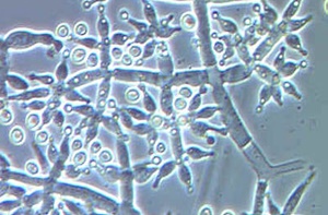
Dans cette étude, 17 souches de Trichoderma on été isolées à partir d’échantillons du sol de différentes origines (champs et forêt d’arganier). Les espèces ont été identifiées par la caractérisation moléculaire des cultures monospores des isolats de Trichoderma et leur potentiel d’antagonisme a été évalué contre trois champignons phyto-pathogènes telluriques (Fusarium oxyxporum, verticillium dahlia et rhizoctonia solani). Après l’extraction de l’ADN, le facteur d’élongation et de traduction tef1 a été utilisé comme marqueur moléculaire pour amplifier, séquencer et par la suite caractériser les 17 souches qui ont été comparées avec leur ex-types déjà référencés. Par conséquent, l’identification moléculaire par tef1 a permis la caractérisation de trois espèces de Trichoderma à savoir T. afroharzianum, et T. guizhouense appartenant à la branche Harzianum, et T. longibrachiatum appartenant à la branche Longibrachiatum. Les 17 isolats candidats ont été sélectionnés par une méthode de criblage basée sur la PCR (polymerase chain reaction). L’évaluation du potentiel antagoniste des souches de Trichoderma contre les champignons phytopathogènes telluriques (F. oxysporum, R. solani et V. dahliae) a été réalisée par la méthode de confrontation sur boîtes de pétrie. L’étude in vitro de l’activité antagoniste par le «Pourcentage of Radial Inhibition Growth» (PRIG %) a permis de suivre l’évolution de la croissance radiale du mycélium des différents champignons phytopathogènes. Le pourcentage le plus élevé (PRIG% = 98%) a été observé pour la confrontation entre l’isolat 8A2.3 et R. solani et le pourcentage le plus faible (PRIG% = 67%) a été observé pour la confrontation entre l’isolat T9i10 et F. oxysporum. D’un autre coté, T9i12 qui est un isolat de l’espèce T. reeseia a engendré une inhibition élevée de la croissance radiale du mycélium des différents pathogènes.


Mots clés: Trichoderma spp, Fusarium oxysporum, Verticillium dahlia, Rhizoctonia solani, champignons antagonistes.

INTRODUCTION

Soil borne pathogens attack a wide range of susceptible plants and cause telluric diseases like seed decay, damping off, root rot and blights (Sneh et al., 1991; El Amraoui et al., 2015).

Some soil borne fungi survive for several years in soil because of their conserved structures then germinate and develop under favorable environmental conditions. In fact, management of diseases caused by soil borne pathogens is difficult. Synthetic fungicides and fumigants, though still used, are not considered a potential solution to sustainable plants production because of their non specific targets application and negative impact on human health and environment (James et al., 1992; Howard et al., 1994).

Trichoderma spp. is a cosmopolitan fungus common in different biotopes and shows different interactions with their neighboring lives. Trichoderma spp. is abundant in soil and involves different kind of interaction with other micro-organisms in the rhizosphere. Some Trichoderma spp. are opportunistic and show parasitic lifestyle against other soil borne fungi what makes them interesting mycoparasitic fungi for the biocontrol of soil borne pathogens. Therefore, many researches in the literature recognized tremendous antagonistic profiles of different Trichoderma spp. against soil borne pathogens. These Trichoderma are used as biological control agents against fungal phytopathogenes (Chet, 1987; Cook, 1993; Papavizas, 1985; Dighton et al., 2005).

Selection of antagonistic candidates in this genus depends on their relevant antimicrobial capacity to reduce either pathogen population or perturb its pathogenic pathway (Rodrigez et al., 2008).

Some Trichoderma species including Trichoderma harzianum, Trichoderma atroviride, T. virens, and Trichoderma asperellum revealed mycoparasitic mode of action. That is, almost all Trichoderma species showed necrotrophic hyperparasitism up on other fungi. The former synthesize chitinases and other proteases that degrade pathogenic fungi cells fed by Trichoderma (Elad and Kapat, 1999; Pozo et al., 2004, Sanz et al., 2005).

Ability of Trichoderma spp. to interact with and antagonize other fungal soil borne pathogens tightly depends on its biodiversity and natural habitat. In fact, Trichoderma species and strains contain tremendous antagonistic profiles due to production of a wide range of secondary metabolites and enzymes. Therefore, each isolate perform specific interaction at the site of living (Hyde, 2005; Suryanarayanan and Hawksworth, 2005).

Trichoderma sp. is known as producer of destructive membrane enzyme (i.e. chitinases) and used as commercial biocontrol agent. However, not all of Trichoderma species are potential antagonists. Therefore, molecular identification of species belonging to Trichoderma genus is mandatory. In fact, molecular markers are widely used during screening of Trichoderma antagonistic candidates to avoid any misleading in identification, monitoring and/or labeling of potential antagonistic candidates (Watts et al., 1988; Schmoll, 2014; Jaklitsch, 2015).

ITS1 and ITS2, tef1 and/or RNA polymerase gene rpb2 are the most used molecular markers in phylogenetic analysis for identification and characterization of Trichoderma species (Jaklitsch, 2006; Jaklitsch, 2011).

In addition, this molecular tool has been developed for high throughput sensitive identification of Trichoderma spp. and considered as a crucial step in early screening of potential antagonists against soil borne pathogens (Kohl et al., 2012; Schmoll, 2010).

Authors in the literature confirmed that molecular approaches led to more refined identification of this genus even at clade and sub-clade level. Tef1 molecular marker is considered sensitive tool for identification, monitoring and labeling of Trichoderma antagonistic candidates during screening procedures. Highly sensitive molecular tools like tef1 marker are highly efficient barcode for Trichoderma species identification regardless confusing morphological characteristics in this genus like color of a colony (Jaklitsch, 2009; Jaklitsch, 2014).

In vitro methods are considered among fast screening techniques and carried out to select Trichoderma antagonistic candidates. For instance, T. asperellum and T. harzianum have been selected as antagonists against different pathogens using dual culture plates. Dual culture plate has been routinely used to select Trichoderma strains with antagonistic activity and omit those who do not present any biological potential (Philion, 1994; Honor, 1996; Matarese, 2012).

In this study, we attempt to select antagonistic candidates among 17 Trichoderma isolates using dual culture assay.

Dual plate test was performed in order to investigate antagonistic potential of Trichoderma isolates. Antagonistic activity was evaluated measuring the capacity of Trichoderma isolates to inhibit mycelium growth of Rhizoctonia solani, Fusarium oxysporum and Verticilium dahlia; common soil borne pathogens of Oak (Halmschlager and Kowalski, 2004; Matarese et al., 2012).

Translation elongation factor alpha 1 (tef1) gene is used as a marker to identify Trichoderma isolates at the species level (Jaklitsch, 2011).

MATERIAL AND METHODS 

Obtaining Trichoderma isolates

Among all fungi isolated from 22 Moroccan soil samples, 17 cultures resembling Trichoderma spp were developed (Table 1).

The strains of F. oxysporum, V. dahlia and R. solani (Table 1) used for the confrontation assays were kindly provided by the Institute of Forest Entomology, Forest Pathology and Forest Protection, University of Natural Resources and Life Sciences, Vienna (BOKU) (IFFF culture collection).

Culture medium

For isolation, cultures were grown on potato dextrose agar medium (PDA, Difco supplemented with ascorbic acid) and were identified by morphological features to genus level.

For genomic DNA extraction and dual culture test, we growed all Trichoderma spp on petri dishes containing Malt extract agar medium which is used to obtain monospore cultures.

Molecular identification of Trichoderma spp. by PCR and sequencing of tef1 DNA

The genomic DNA of Trichoderma strains was extracted according to the fast DNA extraction method of fungi (El Khoury, 2011; Schmoll, 2004). The Trichoderma strains were grown in Malt extract agar at 28°C for 24 hours (hr). Trichoderma mycelium was harvested and added to lysis buffer tubes. Then, the samples were incubated at 65°C for 15 minutes (min), allowed to cool in ice and filled with 150 µL of P3 buffer (3.0 M potassium acetate, pH 5.5) containing potassium acetate, glacial acetic acid and sodium hydroxide. After a full speed centrifugation at 15.000 rpm for 10 min, the supernatant was recovered, transferred to new tubes with 700 µL of isopropanol. Full speed centrifugation for 30 min allowed the recovery of the DNA, the supernatant was discarded and the DNA pellet was washed with ethanol and stored at -20 °C in 50 µL of distilled water (El Khoury, 2011; Schmoll, 2004).

Primers were: EF1-728 5′-AGAGTTTGATCCTGGCTC AG-3’and TEF1-LLE 5’-GGTTACCTTGTTACGACTT-3’ to amplify the fourth and fifth intron and a part of the large exon of translation elongation factor alpha 1 gene. The amplification reaction was performed in a final volume of 10 µL containing 0.3 µM of each primer, 10 mM each dNTP (Jaklitsch, 2011), 0.1 unit Taq DNA polymerase and 1 µL of DNA (30 ng) and 1 × Taq polymerase buffer. The mixture was first denatured at 95 °C for 2 min, then 40 cycles of PCR (Jaklitsch, 2011) were performed with annealing temperature at 55 °C for 30 seconds (sec) and primer extension at 70 °C for 1 min 30 sec. At the end of the last cycle, the mixture was incubated at 70 °C for 3 min. For each reaction, a negative control without DNA template was included. Efficient amplification was confirmed by gel electrophoresis on 1.5% agarose gel. PCR products were purified and sequencing was done at Eurofins Genomics (Ebersberg, Germany). Phylogenetic analyses were performed in MEGA 6 software program using Neighbor Joining distance algorithm method (Jaklitsch, 2009; Saitou and Nei, 1987; Samuels, 2006). Stability of clades was evaluated by bootstrap rearrangement (1000 replicates) displayed next to the branches (Felsenstein, 1985). Only Harzianum clade tree was calculated.

Dual culture test

The Trichoderma strains were tested in vitro for their antagonistic activity against F. oxysporum, V. dahliae and R. solani according to the method of Dickinson and Skidmore (1976). The confrontation assays allowed assessing the capacity of the Trichoderma strains to inhibit the mycelia growth of the above mentioned phytopathogenic fungi.

Malt extract agar (MEA) discs of 6 mm diameter, cut from the edge of an actively growing colony of each antagonist and pathogen, were placed at opposite sides (4.5 cm from each other) on fresh MEA medium plates (Figure 1a). The radii of the developing pathogen’s mycelium were measured in the direction of antagonist’s colony (R1) (Figure 1b) three times a day, until contact. Each antagonist/pathogen combination was set up in triplicate and the inoculated plates were incubated at 24±2 °C with a photoperiod of 12 hr/12 hr darkness/light.

The inhibition of mycelia growth in percent PRIG% (Percentage of Radial Inhibition Growth) was calculated after 14 days using the equation (1) ascribed by Skidmore and Dickinson (1976).

Where, R1: Radius of mycelia growth of pathogenic fungus in control plates (without Trichoderma) in mm.

R2: Radius of mycelia growth of pathogenic fungus in the presence of Trichoderma in mm.

Statistical analysis

Data were processed to calculate basic statistical parameters.

The Percentages of Radial Inhibition Growth PRIG% of pathogens mycelia were subjected to one way ANOVA to compare marginal means between different pathogens, two ways ANOVA was conducted to assess interaction effect between pathogens and Trichoderma isolates combinations. Statistical analysis of only 14 days data is presented (Korsten, 1993; Korsten, 1995; Skidmore and Dickinson, 1976).

RESULTS AND DISCUSSION

Molecular identification of Trichoderma

Trichoderma isolates were identified based on phylogenetic studies of their tef1 gene. Sequences of tef1 gene were submitted to NCBI GenBank database. Comparison of tef1 sequences of isolates to GenBank accessions was performed using nucleotide BLAST alignment program. Sequences alignment revealed similarities with 90 to 99% and 100% homology between tef1 sequences of isolates and Trichoderma harzianum and Trichoderma reesei species respectively.

Phylogenetic analysis of tef1 gene is represented by Harzianum clade tree as illustrated in figure 2.

It appears that, Trichoderma tef1 sequences showed variability among isolates and allowed to obtain distinguishably three Trichoderma species. Distance methods allowed strong resolution in tef1 phylogenetic analysis. tef1 phylogenetic analysis allowed identification and distinction between closely related Trichoderma species in the same clade. Therefore, we obtained three different species; T. afroharzianum and T. guizouhense both belong to Harzianum clade (Figure 3) and reesei species that belong to Longibrachiatum clade.

Previous studies confirmed that Trichoderma phylogenetic analysis using tef1 as molecular marker is more reliable especially for determination of closely related Trichoderma species. Elongation factor alpha gene (tef1) is highly recommended for the reconstruction of phylogenetic trees and identification of Trichoderma/Hypocrea species to avoid misleading characterization (Kubiceck and Druzhini, 2008; Jaklitsch, 2011; Jaklitsch, 2015).

In this study, tef1 phylogenetic analysis allowed allocation of three species according to their habitats distribution (Chaverri et al., 2003; Jaklitsch, 2009; Samuel, 2006).

T. afroharzianum species was isolated from its natural origin in Argania soil in south of Morocco and from Allal tazi soil in North West of the country. However, Trichoderma reesei was isolated exclusively from soils of Argania forest in south. T4.1 isolate corresponding to T. guizouhense species was sampled from Allal tazi soil in North West of the country. Further isolation and molecular identification must be done for more reliable data interpretation related to geographical distribution of Trichoderma species in the country (Jaklitsch 2011; Jaklitsch and Voglmayr, 2015; Kubicek, 2008).

Antagonistic activity of Trichoderma isolates

Dual plate culture method showed considerable antagonistic activity against the three different pathogens when confronting 17 Trichoderma isolates and results of PRIG% means are presented in Table 2.

The Tukey range test (a=0.05) assorted nine distinguishing groups of Trichoderma isolates depending on different PRIG% intervals. First group contains the isolate 8A2.3 with 95.3% as the highest PRIG% mean against three pathogens. Second group contains T3.2 and 8A3.3 reducing the radial mycelia of pathogens by 94.0%, third group contains 8A4.2, 8A1.2, T4.1 and T9i12 by 93.0%, the fourth group T6.1, T9i16, T9i10 by 89.0% - 83.0% PRIG% intervals followed by fifth group T9i9 and T9i8.3 by 82.0% and the sixth group T9i7, T9i11 reducing radial growth of pathogens mycelia by 81.0% - 80.0% respectively. Other orphan groups containing one isolate each are cited as following; T2.1 with 89.9%, group T9i5 with 81.9% and group T9i14 with 77.3%.

Korsten (1995) developed a growth inhibition categories on a scale ranking antagonistic in vitro screening by dual plate culture where 0 scale = no inhibition growth, 1 scale = 1 to 25% inhibition, 2 scale = 25% to 50%, 3 scale = 50% to 75% and 4 scale = 75% to 100%. Therefore, we proposed to scale our Trichoderma isolates using Korsten antagonism scale in Table 2.

Almost all T. afro-harzianum isolates T8A.3.3, T8A.2.3, T8A.4.2, T8A.1, T6.1, T.3.2, T9i14.1, T2.1 et T4.1 showed the highest inhibition percentage compared to the other T. longibrachiatum isolates T9i5, T9i8.3, T9i7, T9i9, T9i10, T9i11, T9i16, with less antagonist performance against the three pathogens with exceptions like T9i12 as shown in Table 2.

Analysis of variance showed differences in inhibition activity assessed by a significant PRIG% means with P = 0.000 ranking F. oxysporum with highest PRIG% means followed by V. dahlia, and by R. solani.

Two ways ANOVA variance analyses show significant interaction P = 0.000 in Trichoderma isolates – pathogen combinations. PRIG% values were statistically significant (P ≤ 0.05) for each Trichoderma isolate confronting different pathogens suggesting that radial growth inhibition of pathogen mycelium depends tightly on the presence of the antagonist Trichoderma (Rahman et al., 2009).

Antagonistic activity was also evaluated by observation of Trichoderma overgrowth up on pathogens and spores production and proliferation in dual plates.

It seems that all Trichoderma colonies overgrow up on pathogens colonies and that was visible after few days of both fungi contact in dual plate. Trichoderma isolates produced spores when confronting pathogens and proliferate once their mycelium cover pathogens mycelium. No color changes have been noticed in Trichoderma cultures (Prisa, 2011).

DISCUSSION

Prior work has documented the effectiveness of many Trichoderma species as biocontrol agents to control plant pathogens thanks to their combined antagonistic mechanisms. For instance, Lorito and co-workers reported that antibiosis is one of the mechanisms of Trichoderma antagonistic candidates. They focused on effect of secondary metabolites and antibiotics production on pathogens growth cease. Other studies focused on mycoparasitic mechanism based on production of degrading enzymes (i.e. chitinases and β-1,3-glucanases) (Chet, 1987; Lorito et al., 2010).

Simple in vitro screening techniques like dual culture plate are used to select varieties of antagonistic profiles of the genus. Dual plate technique is widely used to discover antagonistic potential of Trichoderma spp. and its direct interaction with the pathogen.

Another point, researchers have demonstrated high sensitivity of molecular marker to detect, identify and monitor the fungus of interest during screening procedures. Jaklitsch have recently worked on advanced molecular strategies for reconstruction of Trichoderma clades. Jaklitsch and co-workers have demonstrated that ITS, tef1 and rpb2 are remarkably suitable for Trichoderma identification. They reported that using tef1 alone seems to be satisfactory to discriminate between closely related species of this genus. In the other hand, integrated genetic analysis is crucial for specific characterization of the antagonistic profiles of different species which is a costly process (Jaklitsch, 2006; Jaklitsch, 2011).

Therefore, in this study we tested the effectiveness of tef1 phylogenetic study in identifying Trichoderma isolates. This study compares well with previous tef1 phylogenetic analysis. Furthermore, we attempt to establish a preliminary screening of antagonistic candidates among 17 Trichoderma isolates.

We found that in vitro test resulted in labeling some antagonistic candidates among Trichoderma isolates based on dual culture test. That is, significant high inhibition of pathogens’ mycelium was recorded. Moreover, all 17 Trichoderma isolates performed tropic growth over almost all pathogens mycelium.

It can be inferred that dual culture test is promised selection method of antagonistic candidates during screening procedure. However, it must be applied with cautions. In fact, dual plate is a small esthetic micro-environment that does not show the real behavior of Trichoderma isolates. Therefore, further green houses and field experiments are highly recommended to evaluate antagonistic activity implicating other components.

With tef1 analysis we could label antagonistic candidates among Trichoderma spp. by identifying isolates at the species level. Therefore, Trichoderma harzianum species isolated from Argan forest (i.e. T8A4) were able to inhibit significantly growth of pathogens. However, T. reseei isolates showed less promising antagonistic effect during dual confrontation test except T9i12 reseei isolate. T9i12 inhibits significantly pathogens’ growth.

Other observations for antagonistic evaluation were overgrowth and sporulation over pathogens colony. Trichoderma spp. showed interesting antagonistic interaction like over growth and sporulation over three pathogens.

This finding encourages for more tests based on microscopic observation of short loops, coiling and degradation of pathogens mycelium when in contact with Trichoderma hyphae.

Most notably, Trichoderma spp. inhabiting Moroccan soil; Argania forests, where climate conditions are semi-arid, represent so far an overlooked habitat to isolate Trichoderma spp. Therefore, identifying and classifying Trichoderma species inhabiting new ecological niches like Argania forests may lead us to detect interesting antagonistic candidates (Khattabi, 2004; Druzhinina et al., 2010).

REFERENCES 

Chaverri P. Samuels G. (2003). Trichoderma/Hypocrea (Ascomycota, Hypocreales, Hypocreaceae): species with green ascospores. Studies in Mycology, 48: 1–116.

Chet I. (1987). Trichoderma application, mode of action and potential as biocontrol agent of soil borne plant pathogenic fungi in innovative approaches to plant disease control. John Wiley & Sons, Inc.

Cook R. J. (1993). Making greater use of microbial inoculants in agriculture. Annual Review of Phytopathology, 31: 53–80.

Dighton J., White J.F., Oudemans P. (2005). The fungal community; its organization and role in the ecosystem. Third edition, Taylor & Francis, 93–115.

Druzhinina I.S., Kubicek C.P., Komoń-Zelazowska M., Mulaw T.B., Bissett J. (2010). The Trichoderma harzianum demon: complex speciation history resulting in coexistence of hypothetical biological species, recent agamospecies and numerous relict lineages. BMC Evolutionary Biology, 10: 94–107.

El Amraoui B., Biard J., F., Ikbal F., El Wahidi M., Kandil M., El Amraoui M., Fassouane A. (2015). Activity of Haliscosamine against Fusarium oxysporum f.sp. melonis: in vitro and in vivo analysis. Springer Plus, 4: 1–5

El Khoury A., Atoui A., Rizk T., Lteif R., Kallassy M., Lebhiri A. (2011). Differentiation between Aspergillus flavus and Aspergillu sparasiticus from pure culture and aflatoxin-contaminated grapes using PCR-RFLP analysis of aflR-aflJ intergenic spacer. Food Science, 76: 247–53.

Felsenstein J. (1985). Confidence limits on phylogenies: an approach using the bootstrap. Evolution, 39: 783–791.

Hermosa R., Rubio M.B., Cardoza R.E., Nicolás C., Monte E., Gutiérrez S. (2013). The contribution of Trichoderma to balancing the costs of plant growth and defense. International Microbiology, 16: 69–80.

Honor M. (1996). Screening and development of application techniques for rhizobacteria as biological control agents for green foxtail (Setaria viridis (L.) Bauve). Master thesis of pest Management, SIMON FRASER University, Ontario, Canada, 103 pages: 19–21.

Howard, R. J., Garland, J.A., Scaman, W.L. (1994). Diseases and pests of vegetables crops in Canada. Canadian Phytopathology and Entomology Society, Otawa, Ontario, Page 32.

Jaklitsch W.M. (2009). European species of Hypocrea Part I, the green-spored species. Studies in Mycology, 63: 1–91.

Jaklitsch W.M. (2011). European species of Hypocrea part II: species with hyaline ascospores. Fungal Divers, 48: 1–250.

Jaklitsch W.M., Voglmayr H. (2015). Biodiversity of Trichoderma/Hypocreaceae in southern Europe. Studies in Mycology, 80: 1–87.

Jaklitsch W.M., Samuels G.J., Dodd S.L., Lu B.S. Druzhinina I.S. (2006). Hypocrearufa / Trichoderma viride: a reassessment, and description of five closely related species with and without warted conidia. Studies in Mycology, 56: 137–177.

Khattabi N., Ezzahiri B., Alouali L., Oihabi A. (2004). Antagonistic activity of Trichoderma isolates against Sclerotinum rolfsii: screening of efficient isolates from Morocco soils for biological control. Phytopathologia Mediterranea, 43: 332–340.

Korsten L. (1993). De Villiers E. E., Rowell A., Kotze J. M., (1993). Postharvest biological control of avocado fruit diseases. South African Avocado Growers Association Yearbook, 16: 65–69.

Korsten L., De Jager E.E. (1995). Mode of action of Bacillus subtilis for control of avocado post harvest pathogens. South African Avocado Growers Association Yearbook, 18:124–130.

Kubicek C.P., Mach R.L., Peterbauer C.K. Lorito M., (2001), Trichoderma: from genes to biocontrol. J. Plant Pathology, 83:11–23.

Kubicek C.P., Komon-Zelazowska M., Druzhinina I.S. (2008). Fungal genus Hypocrea /Trichoderma: from barcodes to biodiversity. Journal of Zhejiang University Sciences B, 9: 753–763.

Lorito M., Woo S.L., Harman G.E., Monte E. (2010). Translational Research on Trichoderma: From ‘Omics to the Field. Annual Review Phytopathology, 48: 1–19.

Matarese S., Sarrocco S., Gruber V., Eidl-Seiboth S., Vannacci G. (2012). Biocontrol of Fusarium head blight: interactions between Trichoderma and mycotoxigenic Fusarium. Microbiology, 158: 98–106.

Papavizas G.C. (1985). Trichoderma and Gliocladium: Biology, ecological and potentiality for biocontrol. Annual Review Phytopathology, 23: 23–54.

Philion J. (1994). Screening of potential fungal antagonists of pseudothecial formation by the apple scab pathogen Venlliria inaeqllal, Master of plant science. McGill University, Quebec, Canada, Page 79.

Prisa D., Sarrocco S., Burchi G., Vannacci G. (2013). Endophytic ability of Trichoderma ssp. as inoculants for ornamentals plants innovative substrates. Journal of biocontrol of plant pathogens in sustainable agriculture, IOBC – west palaeartic regional section, 86: 169–174.

Rahman M. A., Begum M. F. , Alam Rahman M. F. (2009). Screening of Trichoderma isolates as a biological control agent against Ceratocystis paradoxa causing pineapple disease of sugarcane. Mycobiology, 37: 277-285.

Saitou N., Nei M. (1987). The neighbor-joining method: a new method for reconstructing phylogenetic trees. Molecular Biology and Evolution, 4: 406-425.

Samuels G.J. (2006). Trichoderma: systematics, the sexual state, and ecology. Phytopathology, 96:195–206.

Sarrocco S., Forti M., Vannacci, G. (2004). Mycoparasitism against sclerotia of Sclerotium rolfsii and Sclerotinia sclerotiorumis widespread within the genus Trichoderma in management of plant diseases and arthropod pests by BCAs and their integration in agricultural systems. Journal of biocontrol of plant pathogens in sustainable agriculture, IOBC – west palaeartic regional section, 27: 375–379.

Schmoll M., Zeilinger S, Mach R.L., Kubicek C.P. (2004). Cloning of genes expressed early during cellulose induction in Hypocrea jecorina by a rapid subtraction hybridization approach. Fungal Genetics and Biology, 41:877–887.

Schmoll M., Seiboth B., Druzhinina I. Kubicek C.P., (2014). Genomics analysis of biocontrol species and industrial enzyme producers from the genus Trichoderma, Fungal Genomics, 2nd Edition. The Mycota XIII, 10: 233–256.

Sinclar J.B., Dhingra O.D., (1995), Basic plant pathology methods, Second edition,Taylor & Francis Group, 448 pages: 217–219.

Skidmore A.M., Dickinson C.H. (1976). Colony interactions and hyphal interference between Septoria nodorum and phylloplane fungi. Transaction of the British Mycological Society, 66: 57–64.

Sneh B., Burpee L., Ogoshi A. (1991). Identification of Rhizoctonia species. The American Phytophatological Society Press, St. Paul, Minnesota, USA, 1-133.

Suryanarayanan T.S., Hawksworth D.L. (2005). Fungi from little explored and extreme habitats. In: Deshmukh S.K., Rai M.K. (eds), Biodiversity of fungi; Their role in human life. Oxford & IBH Publishing Co. Pvt. Ltd., New Delhi, India, 33-48.

Tsahouridou P.C., Thanassoulopoulos C.C. (2001). Trichoderma koningii as a potential parasite of sclerotia of Sclerotium rolfsii Cryptogamie. Mycologie, 22: 289–295.

Watts R., Dahiya J., Chaudhary K., Tauro P. (1988). Isolation and characterization of a new antifungal metabolite of Trichoderma reesei. Plant and Soil, 107: 81–84.